Research in the Cherezov Laboratory focuses on elucidating the structure and function of membrane proteins that play critical roles in human health and disease. The laboratory seeks to uncover the fundamental mechanisms underlying the activity of these proteins and to translate this knowledge into rational strategies for therapeutic development.
A primary research emphasis is on G protein-coupled receptors (GPCRs), which are central regulators of cellular signaling and major targets for drug discovery. To investigate these complex systems, our Laboratory develops and applies advanced structural biology approaches, including cryo-electron microscopy (cryo-EM), micro-electron diffraction (microED), and serial X-ray crystallography, enabling visualization of membrane proteins at high resolution in distinct conformational states.
By integrating structural insights with functional studies, the laboratory aims to advance a deep mechanistic understanding of GPCR signaling and to drive structure-based drug discovery. Through innovation in methodology and a focus on biologically and medically significant targets, the Cherezov Laboratory is building a framework for the rational design of next-generation therapeutics.

Research Highlights

GPCR Structure and Function:

Inhibitory Neurotransmitter Receptor

  • Biological significance: GABAB receptors modulate synaptic signaling in the central nervous system and are implicated in neuropsychiatric conditions ranging from addiction to psychosis.
  • Receptor architecture: GABAB is an obligate class C GPCR heterodimer composed of GB1 and GB2 subunits, each containing an extracellular Venus flytrap (VFT) domain connected to a seven-transmembrane domain (7TM).
  • Structural advance: Four cryo-EM structures of the full-length human GB1–GB2 heterodimer captured distinct functional states: inactive apo, two agonist-bound intermediates, and an active agonist/PAM-bound state.
  • Initial activation step: Agonist binding induces closure of the GB1 VFT domain, initiating large-scale conformational rearrangements within the heterodimer.
  • Transmembrane rearrangement: Activation brings the two 7TM domains into close contact through an interface involving TM6.
  • Signal propagation: These rearrangements produce a lever-like conformational change in the GB2 transmembrane domain, promoting the signaling-competent state.
  • Allosteric modulation: A positive allosteric modulator (PAM) binds at the transmembrane dimer interface and stabilizes the active receptor conformation.
  • Significance: The structures reveal the sequence of conformational changes underlying GABAB receptor activation and positive allosteric modulation, providing a molecular framework for understanding signaling by heterodimeric class C GPCRs.
  • Shaye et al, 2020, Nature 584: 298

Circadian Rhythms Regulators

  • Biological significance: The melatonin receptors MT1 and MT2 are GPCRs that play important roles in regulating and maintaining circadian rhythms.
  • Structural advance: High-resolution structures of human MT1 and MT2 were determined using XFEL serial femtosecond crystallography in complexes with melatonin analogs and approved drugs used for insomnia and depression.
  • Ligand recognition: The structures revealed the architecture of the receptor binding pockets and defined key features of the melatonergic pharmacophore, providing insights into ligand and receptor-subtype selectivity.
  • Unusual ligand-entry pathway: Both receptors contain an occluded orthosteric binding pocket connected to the membrane through a membrane-buried channel, suggesting lateral ligand entry from the lipid bilayer.
  • Subtype-specific feature: MT2 contains an additional potential extracellular ligand-entry pathway, distinguishing it from MT1.
  • Ligand specificity: The unusual access pathway may contribute to the receptors' high selectivity for melatonin over structurally related molecules such as serotonin.
  • Disease-associated variants: The MT2 structure enabled mapping of type 2 diabetes-associated SNPs, revealing a cluster at the protein–membrane interface involving TM1 and TM2 that may influence receptor oligomerization.
  • Significance: The structures provide a foundation for developing subtype-selective melatonin receptor ligands and potentially improved therapeutics for sleep and other melatonin-related disorders.
  • Stauch et al, 2019, Nature 569: 284
  • Johansson et al, 2019, Nature 569: 289

Enigmatic Receptor

  • Biological context: The angiotensin II receptors AT1R and AT2R are key components of the renin–angiotensin–aldosterone system but AT2R has distinct and incompletely understood physiological functions.
  • Structural advance: Crystal structures of human AT2R were determined in complex with an AT2R-selective ligand and an AT1R/AT2R dual ligand.
  • Receptor conformation: Both structures captured AT2R in an active-like conformation.
  • Unusual structural feature: Helix VIII adopts a non-canonical position that stabilizes the active-like receptor while preventing recruitment of G proteins and β-arrestins.
  • Functional implication: This unusual architecture is consistent with the lack of conventional AT2R signaling responses in standard cellular assays.
  • Ligand recognition: Structure–activity relationship, docking, and mutagenesis studies identified key interactions governing ligand binding and receptor selectivity.
  • Significance: The structures provide a molecular basis for understanding the distinct functional and pharmacological properties of AT1R and AT2R and may facilitate the design of selective angiotensin receptor ligands.
  • Zhang et al, 2017, Nature 544: 327

Blood Pressure Regulator

  • Biological significance: The angiotensin II type 1 receptor (AT1R) is a GPCR that plays a central role in blood pressure regulation and is an important target for antihypertensive drugs
  • Structural challenge: Structural characterization of AT1R had been limited by difficulties in obtaining high-quality crystals suitable for conventional synchrotron crystallography.
  • Structural advance: The room-temperature structure of human AT1R bound to the selective antagonist ZD7155 was determined at 2.9 Å resolution using serial femtosecond crystallography (SFX) at an X-ray free-electron laser (XFEL).
  • Ligand recognition: The structure revealed key features of AT1R and identified critical receptor–ligand interactions responsible for ZD7155 binding.
  • Drug-binding analysis: Docking of clinically used angiotensin receptor blockers (ARBs) revealed both shared and distinct binding modes among these antihypertensive drugs.
  • Significance: The results provide a structural framework for understanding AT1R function, antagonist recognition, and structure-based design of improved antihypertensive therapeutics.
  • Zhang et al, 2015, Cell 161: 831

Cell Signaling Switchboard

  • Signaling context:   GPCRs signal primarily through G proteins and arrestins, with arrestin binding blocking G-protein coupling and redirecting signaling toward G-protein-independent pathways.
  • Structural advance:   The structure of a constitutively active human rhodopsin–visual arrestin complex was determined using serial femtosecond X-ray laser crystallography (SFX).
  • Receptor–arrestin interface:   Rhodopsin uses multiple structural elements, including TM7 and helix 8, to recruit and interact with arrestin.
  • Arrestin activation:   Arrestin adopts a pre-activated conformation characterized by an approximately 20° rotation between its N- and C-terminal domains.
  • Intracellular-loop recognition: This domain rotation creates a cleft that accommodates a short helix formed by rhodopsin's second intracellular loop (ICL2).
  • Functional validation: Extensive biochemical and mutagenesis experiments support the receptor–arrestin interactions observed in the structure.
  • Significance: The structure provides a molecular framework for understanding GPCR–arrestin recognition and arrestin-biased signaling and demonstrates the utility of SFX for challenging membrane-protein complexes.
  • Kang et al, 2015, Nature 523: 561

Full-length Structure of a Type 2 Diabetes Target

  • Biological significance: The human glucagon receptor (GCGR) is a class B GPCR that plays a central role in glucose homeostasis and is implicated in type 2 diabetes.
  • Structural advance:  The 3.0 Å crystal structure of full-length GCGR was determined in an inactive conformation using serial femtosecond crystallography.
  • Stalk architecture:  The ECD and TMD are connected by a 12-residue stalk that adopts a β-strand rather than the α-helical conformation observed in the previous isolated TMD structure.
  • Extracellular organization:  The first extracellular loop (ECL1) forms a β-hairpin and interacts with the stalk to create a compact β-sheet structure.
  • Functional validation:   Hydrogen–deuterium exchange, disulfide crosslinking, and molecular dynamics studies indicate that the stalk and ECL1 are important for peptide binding and receptor activation.
  • Significance:  The full-length structure provides new mechanistic insights into ligand recognition and activation of class B GPCRs.
  • Zhang et al, 2017, Nature 546: 259

Pain Relief without Addiction

  • Therapeutic rationale:  Bifunctional μ- and δ-opioid receptor (OR) ligands may provide effective analgesia with fewer side effects than conventional alkaloid opiates.
  • Ligand pharmacology:  DIPP-NH₂ is a bifunctional tetrapeptide that acts as a δ-OR antagonist and μ-OR agonist.
  • Structural approach:  The structure of human δ-OR bound to DIPP-NH₂ was determined using serial femtosecond crystallography.
  • Key structural finding:  The receptor-bound peptide adopts an unusual cis-peptide bond between H-Dmt and Tic. 
  • Molecular insights:  The structure reveals specific receptor–peptide interactions underlying the pharmacological properties of opioid peptides.
  • Significance:  These insights provide a structural framework for developing improved opioid analgesics with potentially reduced adverse effects.
  • Fenalti et al, 2015, Nat Struct Mol Biol 22: 265

GPCRs are Allosteric Machines

  • Background:  GPCR pharmacological responses can be fine-tuned by allosteric modulators, but structural insights into these effects have been limited by the resolution of available GPCR structures.
  • Approach:  The human A2A adenosine receptor was engineered by replacing the third intracellular loop with apocytochrome b562RIL, enabling structure determination at 1.8 Å resolution.
  • Structured waters:  The high-resolution structure revealed 57 ordered water molecules within the receptor, organized into three major clusters.
  • Sodium-binding site:   The central water cluster contains a putative Na⁺ ion coordinated near the conserved Asp2.50.
  • Cholesterol and lipids:  Two cholesterol molecules stabilize helix VI, while one of 23 ordered lipids extends into the ligand-binding pocket.
  • Significance:  The structure provides atomic-level insights into how water, sodium ions, cholesterol, and lipids may contribute to GPCR stability, ligand recognition, and function.
  • Liu et al, 2012, Science 337: 232

First Structure of a GPCR Bound to a Diffusible Ligand

  • Background:  G protein–coupled receptors (GPCRs) constitute the largest family of eukaryotic membrane proteins involved in transmembrane signal transduction but their structural studies are limited because of challenges in GPCR crystallization.
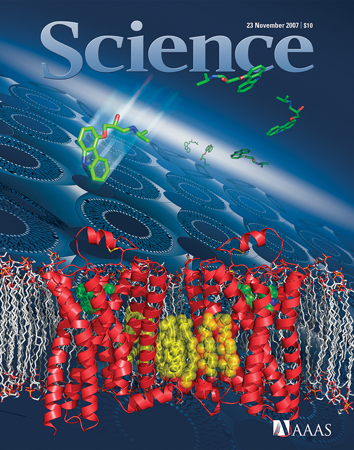
  • Structural advance:  The crystal structure of the human β2-adrenergic receptor (β2AR) fused to T4 lysozyme was determined at 2.4 Å resolution in complex with the partial inverse agonist carazolol using lipidic cubic phase crystallization (LCP).
  • Methodological innovation:  Fusion of T4 lysozyme to β2AR and advancements in LCP crystallization enabled high-resolution structural characterization.
  • Ligand recognition:  The structure provided a high-resolution view of a human GPCR bound to a diffusible small-molecule ligand.
  • Extracellular architecture:  The second extracellular loop (ECL2) is positioned away from the ligand-binding cavity by two closely spaced disulfide bonds and a short α-helical segment, allowing access to the orthosteric pocket.
  • Role of cholesterol:  Cholesterol, which was required for crystallization, mediates a parallel association between β2AR molecules in the crystal lattice.
  • Significance:  The structure provided important insights into GPCR architecture and small-molecule recognition and established a structural framework for studying ligand binding across the GPCR superfamily.
  • Cherezov et al, 2007, Science 318: 1258-65

Structural Biology Methods:

GPCRs Structure Determination by MicroED

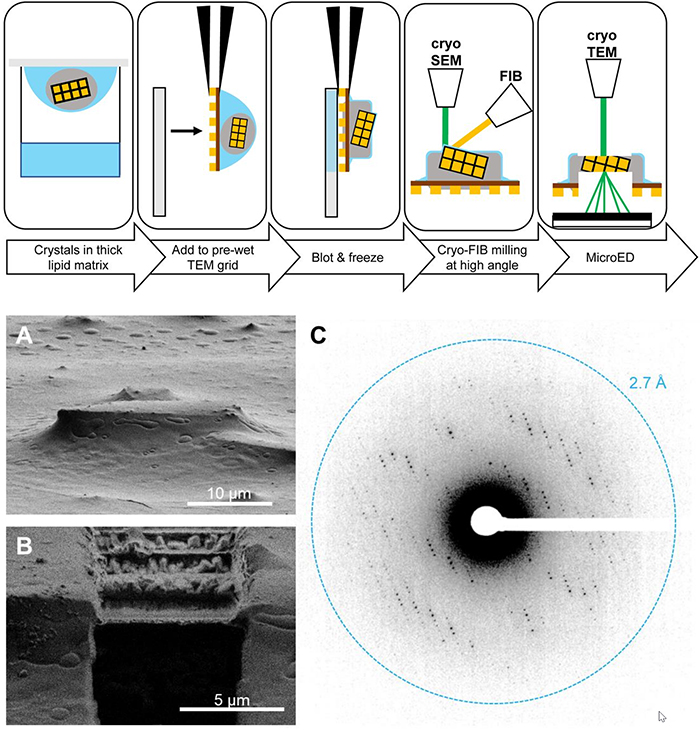
  • Structural challenge:  High-resolution structures of GPCRs without bound signaling partners often require lipidic cubic phase (LCP) crystallization, which typically produces crystals too small for conventional X-ray crystallography.
  • MicroED opportunity:  These GPCR microcrystals are well suited for microcrystal electron diffraction (MicroED), but the viscous, gel-like properties of LCP complicate conventional MicroED sample preparation.
  • Methodological advance:  The study developed an approach that converts LCP into a sponge phase, followed by focused ion-beam (FIB) milling to prepare crystals for MicroED.
  • Proof of concept:  The structure of the human A2A adenosine receptor was determined by MicroED at 2.8 Å resolution.
  • Structural insights:  The structure resolved an antagonist in the orthosteric binding pocket and identified four cholesterol molecules associated with the receptor.
  • Significance:  The method establishes a framework for determining structures of lipid-embedded membrane proteins from individual microcrystals that may be inaccessible to conventional crystallographic approaches.
  • Martynowycz et al, 2021, PNAS 118: 232

Towards SBDD with X-ray Lasers

  • Challenge:   Structure-based drug design (SBDD) benefits from multiple protein–ligand co-crystal structures, but its application to GPCRs has been limited by difficulties in receptor crystallization.
  • Methodological advance:  A rapid approach was developed to determine multiple GPCR–ligand structures using serial femtosecond crystallography (SFX). 
  • Key advantages:  The method eliminates the need for large crystals and requires only submilligram quantities of purified receptor.
  • Proof of concept:  Application to the human β2-adrenergic receptor (β2AR) yielded eight room-temperature co-crystal structures with six different ligands.
  • New structures:  The study provided previously unreported β2AR structures in complex with carvedilol and propranolol.
  • Generality:  The approach was successfully tested with three additional GPCRs, demonstrating its broader applicability.
  • Significance:  Rapid determination of multiple ligand-bound structures can facilitate iterative SBDD and lead optimization for GPCRs and potentially other difficult-to-crystallize membrane proteins.
  • Ishchenko et al, 2019, IUCrJ 6: 106

Serial Femtosecond Crystallography for Membrane Proteins

  • Challenge:  High-resolution crystallography of membrane proteins is limited by difficulties in growing large, well-diffracting crystals and by radiation damage at synchrotron sources.
  •  Approach:  An X-ray free-electron laser (XFEL) with 50-fs pulses was used to minimize radiation damage and collect diffraction data from sub-10-μm microcrystals grown in lipidic cubic phase (LCP).
  • Key result:  The method enabled determination of a high-resolution, room-temperature structure of a human serotonin receptor.
  • Comparison:   The XFEL structure was compared with a conventional synchrotron structure obtained from cryo-cooled crystals approximately two orders of magnitude larger in volume.
  • Structural differences:  The room-temperature structure revealed distinct thermal motions and alternative residue conformations compared with the cryogenic structure.
  • Significance:  Room-temperature serial femtosecond crystallography (SFX) may provide a more physiologically relevant view of GPCR conformational dynamics and structural heterogeneity while enabling structure determination from very small membrane-protein crystals.
  • Liu et al, 2013, Science 342: 1521

Lipidic Cubic Phase (LCP) Technologies

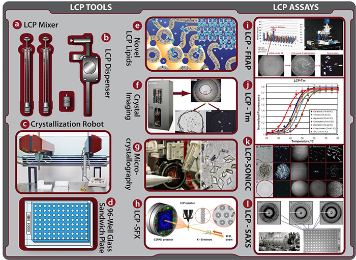
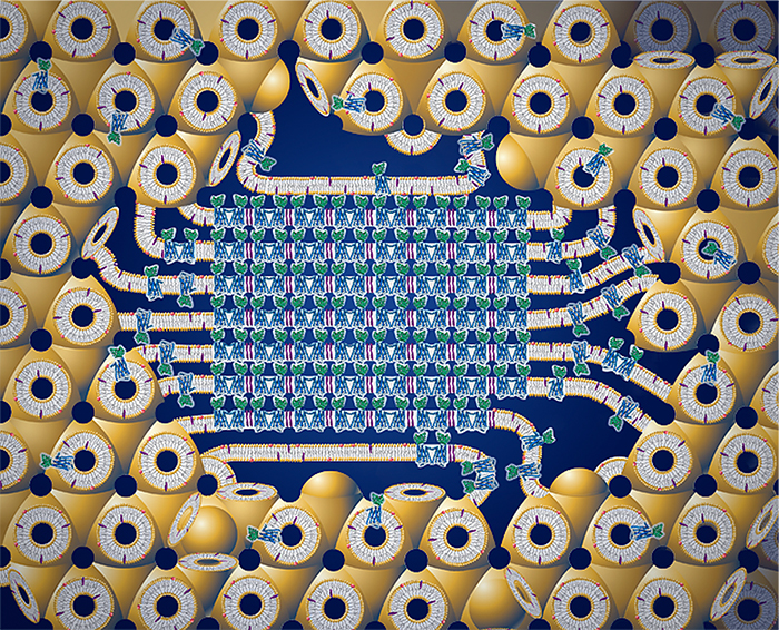
  • LCP as a membrane mimic:  Lipidic cubic phase (LCP) provides a membrane-like lipid environment for stabilizing and crystallizing membrane proteins.
  • Technical challenge:  Broad adoption of LCP methods has been limited primarily by the difficulty of handling highly viscous LCP samples.
  • Recent advances:  Improved pre-crystallization assays, better crystal imaging, and the commercial availability of specialized LCP tools and instrumentation have made the technique more accessible.
  • Demonstrated success:  LCP crystallization has enabled determination of high-resolution structures of challenging membrane proteins, particularly GPCRs.
  • Growing adoption:  These methodological advances and structural successes are encouraging broader use of LCP technologies in membrane protein structural biology. 
  •  Impact:  Wider application of LCP methods should facilitate structural characterization of otherwise difficult-to-crystallize membrane proteins and provide insights into their functional mechanisms and lipid–protein interactions.
  • Cherezov, 2011, Curr Opin Struct Biol 21: 559

Lipidic Cubic Phase Crystallization

  • Method:  A detailed protocol is provided for membrane protein crystallization using lipidic mesophases, also known as the lipidic cubic phase (LCP) or in meso method.
  • Broad applicability:  The method has been successfully applied to diverse membrane proteins, including prokaryotic and eukaryotic proteins, monomers and multimers, α-helical and β-barrel proteins, and proteins with or without chromophores.
  • GPCR applications:  Notable successes include structural determination of the engineered human β2-adrenergic and A2A adenosine receptors.
  • Mesophase preparation:  The protocol describes preparation and characterization of the monoolein-based lipidic mesophase and reconstitution of membrane proteins into LCP.
  • Functional characterization:  Methods are provided to assess protein functionality within the lipidic mesophase before crystallization.
  • Crystallization workflow:  The protocol covers manual setup of LCP crystallization trials and methods for harvesting the resulting microcrystals.
  • Efficiency:  Preparation of the protein-loaded mesophase and manual setup of a crystallization plate can be completed in approximately 1 hour.
  • Significance:  The protocol provides a practical and broadly applicable framework for LCP crystallization of challenging membrane proteins.
  • Caffrey and Cherezov, 2009, Nat Protoc 4: 706